Keratoconus is a progressive eye disease that affects the cornea, the clear front surface of the eye. Over time, the cornea thins and bulges outward into a cone-like shape, leading to blurred vision, irregular astigmatism, and in severe cases, significant visual disability. The disease often begins in adolescence or early adulthood and can worsen gradually over several years.
Understanding the stages of keratoconus is critical because each stage presents unique symptoms, risks, and treatment options. In particular, the early stage plays a decisive role in whether the disease can be managed effectively before it causes severe vision loss.
Let’s explore the progression, classification, and grading of keratoconus, discuss how it evolves from mild to advanced severity, and examine the diagnostic tools and treatments available at each stage. We will also incorporate the latest clinical findings and highlight why early detection is key, especially for Filipinos who may be at higher risk due to genetic and environmental factors.

How Common Is Keratoconus?
Older studies once suggested that keratoconus was rare, with estimates of about 1 in 2,000 people. However, newer diagnostic tools such as corneal tomography have revealed much higher numbers.
- A 2025 PLOS study in Japan found keratoconus in 0.85% of adults and keratoconus-suspect cases in 1.46%.
- Other international research shows prevalence ranging from 1 in 750 to 1 in 50, depending on the population studied and diagnostic methods.
- In the Philippines, while exact prevalence data is limited, eye specialists report increasing numbers of diagnosed cases, likely due to improved screening.
These findings highlight that keratoconus is more common than previously believed, particularly among younger age groups.
Stages of Keratoconus
Early Stage (Mild or Subclinical Keratoconus)
The early stage is the most important to detect, because this is when interventions are most effective.
Key Characteristics:
- Subtle vision changes: mild blurring, ghosting, halos, or double images.
- Increased light sensitivity, especially at night.
- Frequent prescription changes for glasses.
- Corneal shape changes detectable only with imaging tools.
- Sometimes referred to as forme fruste keratoconus or early-onset keratoconus.
Diagnostic Tools:
- Corneal topography is critical, as it reveals irregular curvature before clinical symptoms become severe.
- Pachymetry helps detect thinning, even when patients report only mild symptoms.
Treatment:
- Eyeglasses or soft contact lenses often suffice at this stage.
- Close monitoring is required.
- In some cases, doctors may recommend corneal collagen cross-linking (CXL) to strengthen the cornea and prevent progression.
Intermediate Stage (Moderate Keratoconus)
If the disease progresses, patients move into the moderate stage.
Key Characteristics:
- More pronounced cone-shaped cornea.
- Moderate vision impairment not fully correctable with glasses.
- Worsening astigmatism.
- Corneal thinning becomes more evident.
Diagnostic Tools:
- Keratometry shows increased curvature.
- Tomography can highlight changes in the posterior corneal surface.
Treatment:
- CXL is highly recommended to stop or slow progression.
- Specialty contact lenses such as rigid gas permeable (RGP), hybrid, or scleral lenses may provide clearer vision.
- Studies confirm that early intervention with CXL can stabilize the disease in up to 90% of cases.
Advanced Stage (Severe Keratoconus)
In severe cases, keratoconus reaches its advanced stage, significantly affecting quality of life.
Key Characteristics:
- Severe vision loss.
- Significant corneal bulging.
- Corneal scarring that further distorts vision.
- Complications such as corneal hydrops, where fluid suddenly enters the cornea and causes swelling.
Diagnostic Tools:
- Advanced imaging may no longer provide accurate measurements if scarring is extensive.
Treatment:
- Corneal transplant (keratoplasty) is often required when vision cannot be corrected by lenses.
- Alternatives such as Intacs (intracorneal ring segments) may help in select cases.
- Scleral lenses may still improve vision for some patients.
Treatment Pathways by Stage
Early Stage:
- Glasses, soft lenses.
- Observation and regular monitoring.
- Preventive cross-linking in select cases.
Intermediate Stage:
- Corneal collagen cross-linking (gold standard).
- Specialty lenses: RGP, scleral, hybrid.
Advanced Stage:
- Corneal graft (keratoplasty).
- Intacs or corneal implants.
- Custom scleral lenses.

Prognosis and Long-Term Outlook
The natural course of keratoconus usually spans 10–20 years, often stabilizing by the late 30s or early 40s. However, some patients continue to progress even beyond that age.
Quality of life can be significantly affected. Research from the U.S. estimates that patients with keratoconus may face an economic burden of around $25,000 in lifetime costs, due to frequent eye care visits, specialty lenses, and potential surgery.
For Filipinos, where regular eye check-ups may not always be prioritized, this highlights the importance of early detection and proactive care.
Conclusion
Keratoconus is a disease that follows a clear pattern of progression—from mild, early stages that are often easy to miss, to advanced stages that can cause severe visual disability. The key to protecting vision lies in recognizing the first signs, undergoing proper diagnostic imaging, and seeking treatment before the disease worsens.
By understanding the classification, grading, and treatment pathways, patients and doctors can work together to slow or even halt the evolution of keratoconus.
By submitting this form, you consent to the processing of your personal data in accordance with our Data Privacy Notice.
For those in the Philippines, it is vital to consult trusted eye care providers who have the latest diagnostic tools and treatment expertise. Shinagawa Lasik & Aesthetics, one of the world’s leading LASIK providers, offers comprehensive eye exams, advanced diagnostic imaging, and modern treatments such as corneal collagen cross-linking. With their expertise, patients can feel confident in receiving the care they need to safeguard their vision—before keratoconus reaches its severe stages.
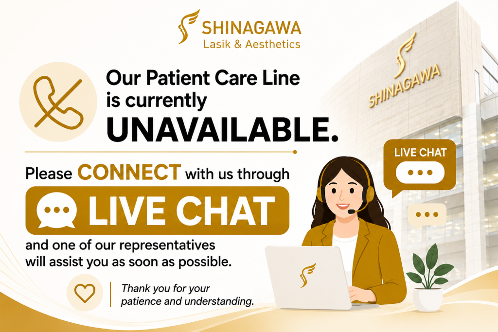
For inquiries, questions, and appointments, call our Patient Care Lines:
🖥 Talk to our Consultants via Livechat: https://direct.lc.chat/6329011/
📱 Instagram: https://instagram.com/shinagawa_ph/
Citations and Resources
Shinagawa LASIK & Aesthetics strives to provide accurate and reliable information regarding LASIK procedures and eye health. We utilize primary sources to support our content, including peer-reviewed scientific studies, data from reputable medical organizations, and expert opinions. We also reference established publications and research where appropriate.
Our commitment to evidence-based information ensures that you receive trustworthy and up-to-date details to make informed decisions about your eye care.
Resources Used in This Article
- PLOS. “Prevalence of keratoconus and keratoconus suspect, and their characteristics on corneal tomography in a population-based study, https://journals.plos.org/plosone/article?id=10.1371/journal.pone.0308892“
- Philippine Journal of Ophthalmology. “Clinical Profile of Keratoconus Patients at the Philippine General Hospital, https://paojournal.com/index.php/pjo/article/view/17/422“
- National Library of Medicine. “Corneal Topography, https://www.ncbi.nlm.nih.gov/books/NBK585055/“
- Science Direct. “Corneal collagen cross-linking: A reviewEntrecruzamiento del colágeno corneal: Revisión, https://www.sciencedirect.com/science/article/pii/S1888429613000824“
- American Academy of Ophthalmology. “What Is Keratoconus?, https://www.aao.org/eye-health/diseases/what-is-keratoconus“
- HMP Global. “Keratoconus continues to progress in some older patients, https://www.hmpgloballearningnetwork.com/site/pophealth/content/keratoconus-continues-progress-some-older-patients“
- Research Gate. “Prevalence and Economic Burden of Keratoconus in the United States, https://www.researchgate.net/publication/375521720_Prevalence_and_economic_burden_of_Keratoconus_in_the_United_States“