
Keratoconus is a progressive eye condition that causes the cornea—the clear, dome-shaped surface at the front of the eye—to thin and bulge into a cone-like shape. This distortion affects how light enters the eye, leading to blurred or distorted vision.
While keratoconus is often discussed as a single condition, specialists actually classify it into different types and stages, based on the shape of the cornea, location of the bulge, and disease severity.
Understanding these classifications is essential because each type influences the choice of treatment, from corrective lenses to advanced corneal procedures such as corneal cross-linking (CXL).
This article explores the types of keratoconus, their characteristics, how they are diagnosed, and why proper classification matters in protecting your vision.
What “Type” Means in Keratoconus
When ophthalmologists talk about the type of keratoconus, they often refer to two major categories of classification:
- Morphological Type – based on the shape and location of the cone on the cornea (e.g., central, inferior, or widespread).
- Clinical or Severity Stage – based on how advanced the disease is, often measured through corneal curvature, thickness, and visual function.
In simpler terms, the morphological type tells us what the cornea looks like, while the clinical classification tells us how far the disease has progressed. Both are vital for accurate diagnosis and treatment planning.
Clinical Classification: Measuring Disease Severity
Beyond shape and size, keratoconus is also classified based on its severity and progression. Eye specialists use two main systems: Amsler-Krumeich and Belin ABCD.
Amsler-Krumeich Classification
The Amsler-Krumeich (AK) system is the traditional grading method, based on parameters like:
- Corneal curvature (measured in diopters)
- Corneal thickness (in micrometers)
- Refractive error and visual acuity
- Presence of corneal scarring
It divides keratoconus into four stages—from mild (Stage I) to severe (Stage IV). However, the AK system primarily evaluates the front surface of the cornea, making it less sensitive in detecting early or subclinical disease.
Belin ABCD Grading System
The Belin ABCD system is a modern classification using tomography-based metrics, allowing a more detailed assessment:
- A – Anterior curvature
- B – Posterior curvature
- C – Corneal thickness at the thinnest point
- D – Best corrected distance visual acuity
This system helps identify early keratoconus before it affects vision, making it invaluable in monitoring disease progression and planning cross-linking treatment.
By integrating anterior and posterior elevation data, the Belin system offers a three-dimensional understanding of corneal changes—something older systems couldn’t do.
How Keratoconus Type Affects Treatment
Each keratoconus type responds differently to various treatments. Management focuses on improving vision and halting disease progression.
Non-Surgical Management
For mild to moderate cases:
- Eyeglasses may help initially but are often insufficient as the disease progresses.
- Rigid Gas Permeable (RGP), Hybrid, and Scleral lenses provide customized optics for irregular corneas.
- Scleral lenses, in particular, vault over the cornea to create a smooth optical surface, improving both comfort and vision.
Corneal Cross-Linking (CXL)
- Recommended when imaging shows active progression (increasing curvature or thinning).
- Uses riboflavin (vitamin B2) and UV light to strengthen corneal fibers, stabilizing the shape.
- CXL is most effective in early to moderate stages and can prevent the need for corneal transplantation.
Surgical Treatments
For advanced keratoconus or severe corneal distortion:
- Intrastromal Corneal Ring Segments (ICRS or Intacs): Small implants inserted to flatten and regularize the cornea.
- Corneal Transplant (Keratoplasty): Necessary in cases of extreme thinning, scarring, or vision loss.
These options are usually determined by your cone type, disease stage, and corneal thickness.
FAQs: What People Also Ask
Yes. As keratoconus progresses, a small nipple cone can expand and evolve into an oval or globus pattern. Regular topographic or tomographic scans help track these changes accurately.
Not directly. While shape influences lens fitting, treatment depends more on progression and corneal stability. For instance, cross-linking (CXL) is recommended once measurable progression is detected, regardless of the cone’s shape.
Yes, it often runs in families and is linked with connective tissue disorders like Ehlers-Danlos or Down Syndrome. However, environmental factors such as eye rubbing and allergies also contribute.
There is no permanent cure, but treatments like cross-linking, specialty contact lenses, and corneal surgery can dramatically improve vision and stop the condition from worsening.
Protecting Your Vision: The Importance of Accurate Diagnosis
Early detection of keratoconus—especially in its forme fruste or subclinical stages—is the key to preserving long-term vision.
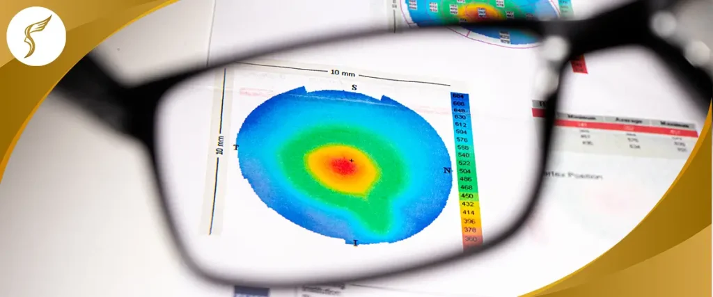
Routine corneal mapping allows doctors to identify microscopic changes before symptoms appear, enabling preventive treatment such as CXL at the right time.
In the Philippines, advanced eye centers now use Japanese-standard tomography and diagnostic tools to classify keratoconus accurately. This ensures every patient receives personalized, stage-appropriate care.
Conclusion
Understanding the types and stages of keratoconus empowers you to take proactive steps toward protecting your vision. Whether you’re dealing with early indicators like forme fruste keratoconus or more advanced patterns such as oval or globus cones, early diagnosis and proper treatment make a significant difference in preserving your eyesight.
At Shinagawa Lasik & Aesthetics, we combine Japanese-standard corneal diagnostics and world-class expertise to detect keratoconus at any stage. Our specialists offer personalized treatments—from corneal cross-linking (CXL) for stabilization to advanced vision correction solutions—ensuring every patient receives precise and effective care.
By submitting this form, you consent to the processing of your personal data in accordance with our Data Privacy Notice.
If you’re experiencing blurry, distorted, or fluctuating vision, or suspect early keratoconus, don’t wait for your symptoms to worsen.
For inquiries, questions, and appointments, call our Patient Care Lines:
🖥 Talk to our Consultants via Livechat: https://direct.lc.chat/6329011/
📱 Instagram: https://instagram.com/shinagawa_ph/
Citations and Resources
Shinagawa LASIK & Aesthetics strives to provide accurate and reliable information regarding LASIK procedures and eye health. We utilize primary sources to support our content, including peer-reviewed scientific studies, data from reputable medical organizations, and expert opinions. We also reference established publications and research where appropriate.
Our commitment to evidence-based information ensures that you receive trustworthy and up-to-date details to make informed decisions about your eye care.
Resources Used in This Article
- PubMed. “Global Incidence and Prevalence of Keratoconus: A Systematic Review and Meta-Analysis, https://pubmed.ncbi.nlm.nih.gov/40833011/”
- BMC Ophthalmology. “Diagnosis of forme fruste keratoconus with scheimpflug photography in Ghanaian patients, https://bmcophthalmol.biomedcentral.com/articles/10.1186/s12886-024-03563-x”
- IOVS. “Differentiation of forme fruste keratoconus from normal cornea using parameters of corneal tomography, aberration, and biomechanics, https://iovs.arvojournals.org/article.aspx?articleid=2269150″
- ResearchGate. “Chasing the suspect: Keratoconus, https://www.researchgate.net/publication/26317902_Chasing_the_suspect_Keratoconus”
- PubMed. “ABCD: A new classification for keratoconus, https://pmc.ncbi.nlm.nih.gov/articles/PMC7856970/”
- ResearchGate. “Management of keratoconus: an updated review, https://www.researchgate.net/publication/371736553_Management_of_keratoconus_an_updated_review”
- PAOJournal. “Clinical Profile of Keratoconus Patients at the Philippine General Hospital, https://paojournal.com/index.php/pjo/article/view/17/422″