
Keratoconus is a complex eye condition that changes the way we see the world—literally. It gradually thins and bulges the cornea into a cone-like shape, leading to distorted and blurred vision. While modern treatments such as Corneal Collagen Cross-Linking (CXL) can halt its advancement, one of the most common questions patients ask is:
“At what age does keratoconus stop progressing?”
The short answer: many patients experience natural stabilization between their 30s and 40s, but this is not guaranteed. The disease’s course depends on age, genetics, and lifestyle factors such as eye rubbing or allergies. Let’s explore how and why keratoconus behaves differently over time—and what you can do to protect your vision.
Understanding the Natural Course of Keratoconus
Keratoconus usually begins during adolescence or early adulthood, when the cornea’s collagen structure is still flexible. The condition often progresses quickly during these years, causing noticeable changes in vision prescriptions and corneal shape.
The natural history of the disease suggests that as we age, the cornea undergoes biological stiffening due to age-related collagen cross-linking—a process similar to how the skin becomes less elastic over time. This gradual increase in corneal rigidity helps stabilize the shape of the cornea, reducing the rate of progression.
However, “natural stabilization” doesn’t mean the disease completely stops. Some individuals—especially those with chronic allergies or vigorous eye rubbing habits—may still experience mild deterioration even after their forties. This means that keratoconus is not self-limiting for everyone.
Timeline Overview:
- Teens to early 20s: Onset and rapid progression
- 30s: Possible slowing or plateauing phase
- 40s and beyond: Often more stable, but continued monitoring is essential
How Doctors Determine if Keratoconus Has Stopped Progressing
Stability in keratoconus is determined not by how long you’ve had it, but by what diagnostic tests show over time.
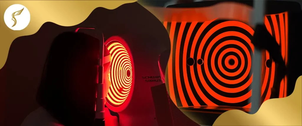
Ophthalmologists rely on sequential imaging and objective assessments to compare the cornea’s shape and thickness across months or years.
The main tools include:
- Corneal Topography and Tomography (like Pentacam or Orbscan) to map curvature changes
- Kmax (Maximum Keratometry): measures the steepest point on the cornea
- Thinnest Corneal Thickness (TCT): tracks thinning progression
- Posterior Corneal Elevation: detects subtle bulging not visible on the surface
If these measurements show no significant change—typically less than 1 diopter increase in Kmax over a 12–24 month period—the condition is considered clinically stable.
That stability often goes hand-in-hand with refractive stability (no worsening of vision or prescription).
In short: When your corneal metrics remain steady over time and your vision stops changing, doctors consider the keratoconus to have reached a stable phase.
At What Age Does Keratoconus Usually Stabilize?
Research indicates that most patients experience a stabilization threshold between their mid-thirties and early forties, sometimes referred to as the fourth decade of life.
This is the period when natural corneal stiffening from biochemical collagen cross-linking and UV exposure occurs.
Yet, several clinical studies and long-term patient follow-ups have found exceptions. Some individuals—particularly those with pediatric keratoconus, Down Syndrome, or severe atopy (eczema, asthma, allergic rhinitis)—may continue to deteriorate well into their 40s or 50s.
For these cases, relying on natural stabilization alone isn’t safe. The disease’s biomechanical properties vary per person, meaning regular eye check-ups are the only way to ensure true stability.
Why Some People Keep Progressing Beyond 40
While aging typically strengthens the cornea, certain risk factors can override this natural defense mechanism.
- Eye Rubbing: Constant mechanical friction triggers microtrauma and an inflammatory cascade, weakening the corneal structure.
- Allergic Conditions: Chronic atopy—including allergic rhinitis, asthma, or eczema—can make you rub your eyes more often, accelerating thinning.
- Genetic Predisposition: Disorders like Ehlers-Danlos Syndrome or Down Syndrome reduce collagen quality, increasing the risk of continued progression.
- Hormonal Changes: Certain life stages, such as pregnancy, may influence corneal biomechanics.
Patients with a younger age at diagnosis, rapid deterioration, or non-compliance with follow-up care often show more aggressive keratoconus patterns.

How Corneal Cross-Linking (CXL) Stops Keratoconus Progression
When natural stability is not enough, Corneal Collagen Cross-Linking serves as a clinical intervention to force disease cessation.
This procedure strengthens the cornea by saturating it with riboflavin (vitamin B2) and exposing it to controlled UV light, creating new collagen bonds.
There are three main variations:
- Epi-Off CXL (Dresden Protocol): the standard approach; most effective for rapid cases.
- Epi-On or Transepithelial CXL: gentler, suitable for thinner corneas.
- Accelerated CXL: shorter UV exposure but similar stabilization results.
CXL works by increasing corneal rigidity and stiffness modulus, halting progression in both pediatric and adult cases.
In clinical studies, more than 90% of patients achieved stability after CXL, with effects lasting up to a decade or more.
If keratoconus is already advanced, other interventions—such as Intacs (ICRS) or Keratoplasty (corneal transplant)—may be considered for vision restoration.
Conversational FAQs
Most patients see a slowdown between 30 to 40 years old, when corneal stiffening naturally increases. But each case is unique—some continue to progress even after this age.
Yes, though less common. Eye rubbing, genetic factors, and hormonal changes can still influence progression beyond 40.
If your Kmax change is ≤1 diopter over a year or more, with no significant corneal thinning, it’s generally considered stable.
CXL provides long-term stability in most patients, but follow-up exams remain necessary to detect rare re-progression.
No. Contact lenses correct vision distortion but don’t prevent structural corneal changes. Only CXL can halt progression.te
Myths and Facts About Age and Keratoconus
- Myth: Keratoconus always stops by age 30.
Fact: It often stabilizes between 30–40, but some patients continue to progress beyond that.
- Myth: Once stable, it can’t come back.
Fact: Stability doesn’t mean permanent cessation—environmental and mechanical factors can still trigger changes.
- Myth: LASIK can treat keratoconus.
Fact: LASIK is contraindicated for keratoconus, as it can worsen corneal thinning. Treatments like CXL are safer alternatives.
What This Means for You
If you’re in your teens or twenties and recently diagnosed, your keratoconus may still be in its active phase.
If you’re in your 30s or older, your corneas might naturally stiffen, slowing down progression—but only regular tomography scans can confirm this.
Regardless of age, early diagnosis and timely treatment remain key to preserving good vision and avoiding corneal transplant.
Practical Takeaway:
- Don’t assume your keratoconus has stopped just because your vision feels stable.
- Follow your doctor’s recommended imaging schedule.
- Address allergies and eye rubbing habits early to protect your cornea’s long-term health.
Conclusion: Your Eyes Deserve Expert Care
Keratoconus may stabilize with age, but its behavior is unpredictable. Understanding the disease’s biomechanics, tracking your corneal metrics, and seeking timely treatment are essential to maintain stable, clear vision.
At Shinagawa Lasik & Aesthetics, our specialists use Japanese-standard diagnostic technology to monitor corneal changes with precision. Whether you’re seeking early detection, follow-up evaluation, or Corneal Cross-Linking (CXL) treatment, our team ensures you receive care tailored to your condition and lifestyle.
By submitting this form, you consent to the processing of your personal data in accordance with our Data Privacy Notice.
Take control of your eye health—schedule a comprehensive screening at Shinagawa Lasik & Aesthetics, the Philippines’ trusted leader in advanced eye care and vision correction.
For inquiries, questions, and appointments, call our Patient Care Lines:
🖥 Talk to our Consultants via Livechat: https://direct.lc.chat/6329011/
📱 Instagram: https://instagram.com/shinagawa_ph/
Citations and Resources
Shinagawa LASIK & Aesthetics strives to provide accurate and reliable information regarding LASIK procedures and eye health. We utilize primary sources to support our content, including peer-reviewed scientific studies, data from reputable medical organizations, and expert opinions. We also reference established publications and research where appropriate.
Our commitment to evidence-based information ensures that you receive trustworthy and up-to-date details to make informed decisions about your eye care.
Resources Used in This Article
- John Hopkins Medicine. “Keratoconus, https://www.hopkinsmedicine.org/health/conditions-and-diseases/keratoconus”
- The Ophthalmologist. “Busting Keratoconus Myths, https://theophthalmologist.com/issues/2021/articles/jun/busting-keratoconus-myths”
- PubMed. “Effect of ageing on keratoconic corneas, https://pmc.ncbi.nlm.nih.gov/articles/PMC4812003/”
- ELZA Institute. “Study Shows Keratoconus Progression Continues in Older Adults, https://www.elza-institute.com/study-shows-keratoconus-progression-continues-in-older-adults/”
- Frontiers. “Medium to long term follow up study of the efficacy of cessation of eye-rubbing to halt progression of keratoconus, https://www.frontiersin.org/journals/medicine/articles/10.3389/fmed.2023.1152266/full”
- Nature. “Longitudinal assessment of the progression of severe keratoconus based on corneal topography, https://www.nature.com/articles/s41598-024-70084-4″